

Admissions
Explore application requirements for the M.D. program and the Ph.D. in Basic Medical Sciences.
Learn More
Departments
Discover the Division of Medical Education and our basic sciences and clinical departments.
Learn More
Research
Learn about the diverse research opportunities available to students, faculty, residents and fellows.
Learn More
Our mission is to provide innovative research, education, and clinical care in a culture of excellence, discovery, and scholarship to the next generation of physicians, scientists, and educators to advance patient-centered and equitable healthcare.
As a member of the Association of American Medical Colleges (AAMC), the Frederick P. Whiddon College of Medicine delivers comprehensive, accredited education across the medical training continuum. The College offers a Doctor of Medicine (M.D.) program accredited by the Liaison Committee on Medical Education (LCME) and a Ph.D. program in Basic Medical Sciences through the University of South Alabama Graduate School. USA Health residency and fellowship programs are affiliated with the College and accredited by the Accreditation Council for Graduate Medical Education (ACGME).
The future of medical education and research
Construction of a 295,000-square-foot Whiddon College of Medicine building is under way and will bring research and education together under one roof. The new facility, scheduled to open in 2027, will provide a 21st century, interactive learning environment for our students and faculty and foster best practices in teaching the next generation of physicians and scientists.

Learn more
The 2024-2027 COM Strategic Plan [PDF]
The 2023-2024 Annual Report [PDF]
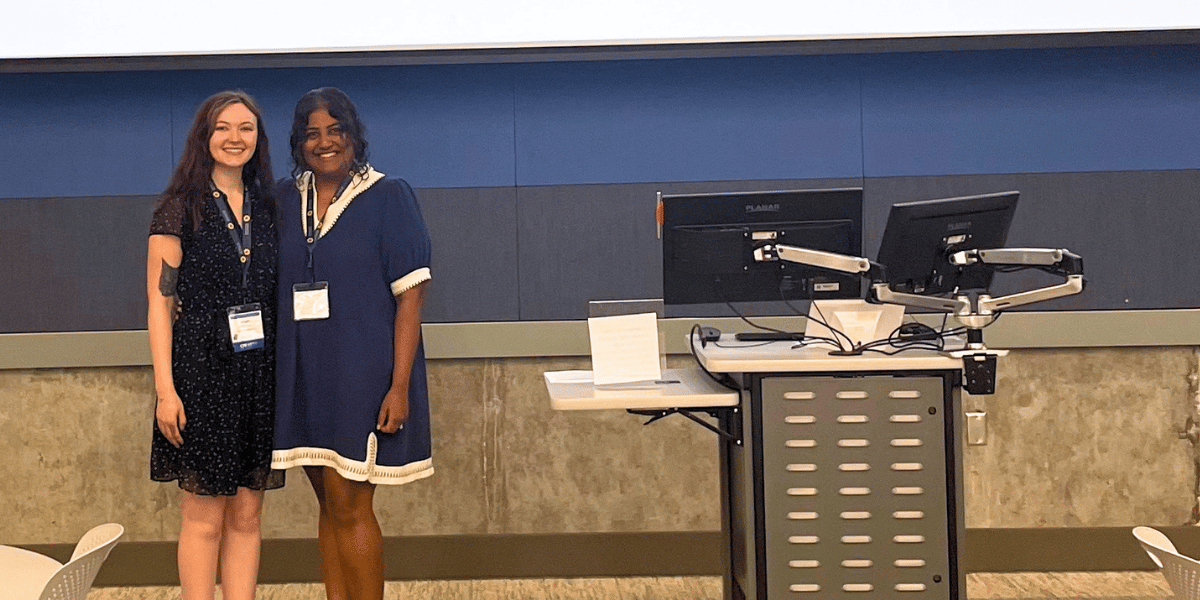 Graduate student gives invited talk at Midwest DNA Repair Symposium
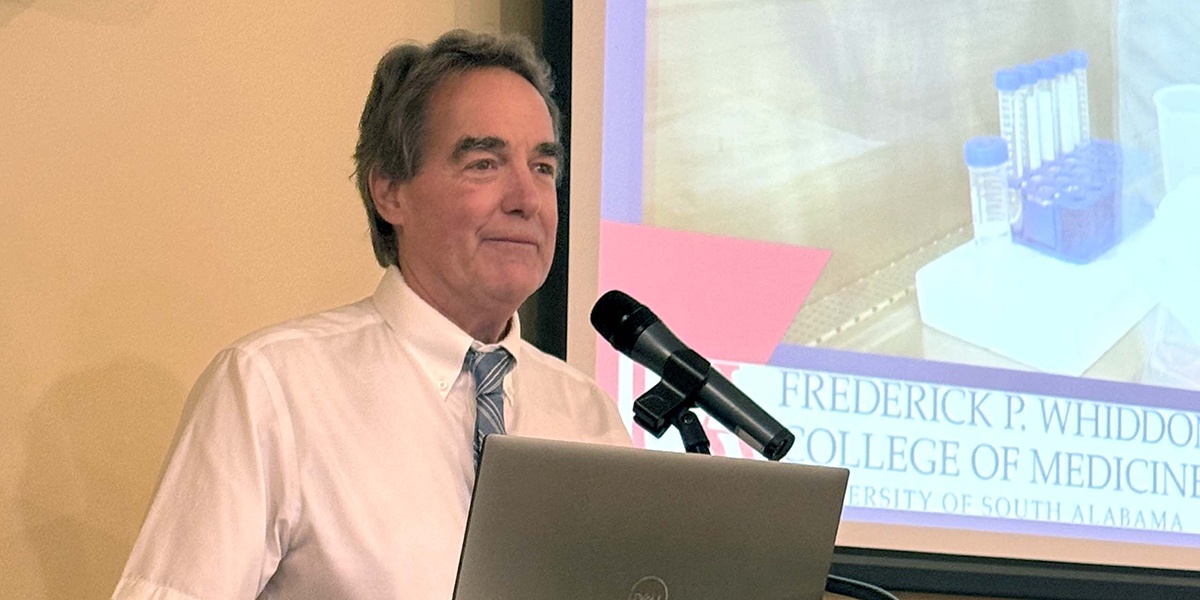 Honkanen retires after transformative career in research and mentorship
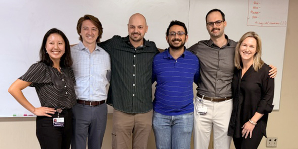 Emergency medicine resident physicians take part in an academic research day