
Instruments
| Description & Location |
|---|
| BD FACSAria-SORP The FACSAria-SORP cell sorters are capable of 12 fluorescence parameters. The sorter has 4 laser lines, including a 60 mW 350 nm UV line, 100 mW 488 nm, 100 mW 561 nm and 40 mW 640 nm lines. The sorters can be used to isolate cells in multiple formats, including 4-way tube sorting and 96-well plate sorting via the Automated Cell Deposition Unit. Location: USA COM Medical Sciences Building & Mitchell Cancer Institute 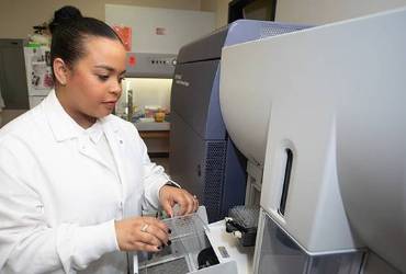 |
| Agilent NovoCyte Opteon High-performance spectral flow cytometer designed for complex, multi-parametric cell analysis. Launched in 2024, it integrates advanced spectral technology into the user-friendly NovoCyte platform, enabling the simultaneous detection of over 40 markers. Location: Mitchell Cancer Institute (MCI) |
| Agilent NovoCyte Quanteon Provides an expanded set of capabilities that accommodate today’s high-end and increasingly sophisticated multi-color flow cytometry assays. You now have the flexibility to choose from up to 25 fluorescence channels utilizing up to four lasers with up to 25 independent detectors. The NovoSampler Q, which can be integrated into different laboratory automation platforms, efficiently processes both FACS tubes (using a a 40-tube rack) and 24-, 48-, 96-, and 384-well plates. The intuitive and industry-leading NovoExpress software has been further advanced, providing an exceptional user experience in data acquisition, analysis, and reporting. Location: USA COM Medical Sciences Building (MSB) 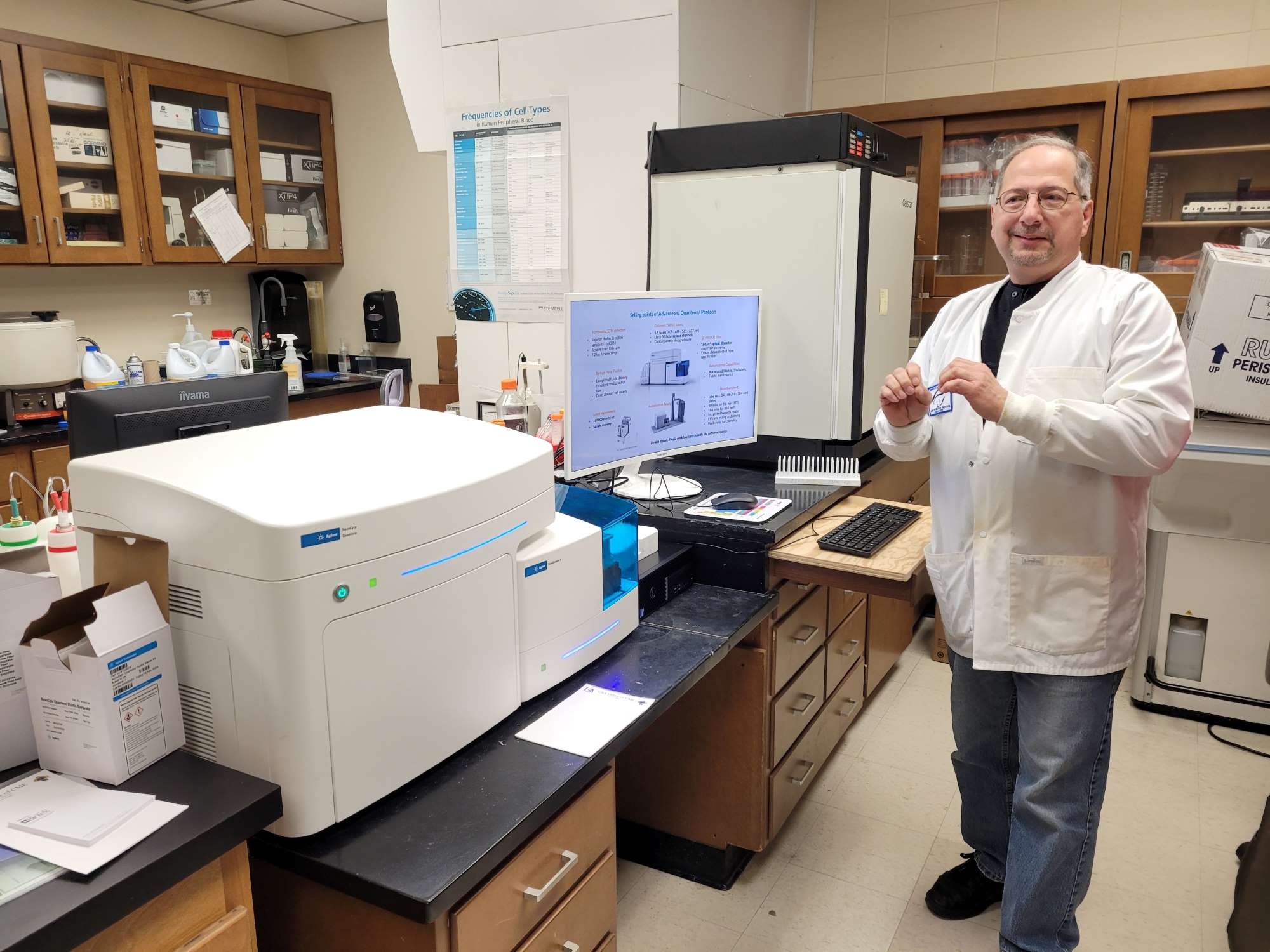 |
| Nexcelom CeligoS microplate based imaging cytometer The CeligoS imaging cytometer is designed for performing assays directly in multi-well plates (6-384 wells). There are many advantages to this approach, including lower demand for cells (allowing for the use of rare primary cells isolated from patient samples) and reagents; as well as analyzing the cells in their native state without the need to enzymatically remove them from their growth substrate, as is required for traditional flow cytometry. It can image cells in Brightfield and 3 fluorescent channels: blue (377/50nm Ex 470/22nm Em) Green (483/32 Ex 536/40 Em) and orange/red (531/40 Ex 629/53 Em). Using advanced software based image recognition there are over 10 specific analysis modes for a wide variety of applications. The CeligoS is also a self-serve instrument after training is provided. Location: Mitchell Cancer Institute (MCI) 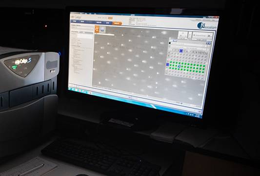 |
| Celsee Prep100 CTC isolation system The Celsee Prep100 uses size exclusion microfluidic filtration to capture CTC from whole blood using a label free method. The system is based on disposable microfluidic chips and this base model employs manual processing to rapidly isolate very rare cells from blood (100-1000 in 10ml of whole blood). Location: Mitchell Cancer Institute (MCI) |
| On-Chip Biotechnologies Microfluidic Cell Sorter (3 laser 6 color) The On-Chip is the world’s first in its class microfluidic cell sorter, developed in Japan. USA MCI was one of the first centers in America to obtain this new technology. Benefits over traditional sorting include the ability to analyze extremely low starting sample volumes/number of cells. The sorting is much gentler and can accommodate a wide range of particles from exosomes to cell spheres using two different size microfluidic chips. The sorter is aerosol free and placed in a BSL2 cabinet allowing for the safe isolation of cells infected with lenti virus or other pathogens. The 3 excitation lasers (405nm, 488nm & 561nm) are collinear so fluorochromes excited by any laser can be detected in any of these channels: FL1(445/20 nm), FL2(543/22 nm), FL3(591.5/43 nm), FL4(607/36 nm), FL5(676/37 nm), FL6(732/68 nm). Location: Mitchell Cancer Institute (MCI) |
| On-Chip Biotechnology SPiS The SPiS companion instrument does single cell or sphere deposition into 96 or 384 well plates in a far more gentle and reliable manner than traditional sorters. Location: Mitchell Cancer Institute (MCI) |
| The ZetaView® TWIN - NTA Nanoparticle Tracking - Video Microscope PMX-220 The 2-laser platform (488 & 640 nm) was installed in the core lab in May 2019. Nanoparticle Tracking Analysis (NTA) captures the Brownian motion of each particle in the video. Based on the different diffusion movements of large and small particles in the surrounding liquid, the hydrodynamic diameter of the particles is determined. Pattern parameters, such as intensity fluctuations, surface geometry and shape of the particles as well as particle concentration are documented at each recording and can be used to distinguish sub-populations. In addition, the charge state of the particle surface (zeta potential) can be measured via the movement of the particles in an applied electric field. Depending on the type of sample and the measuring mode, the measuring range is between 15 nm and 5 μm. Location: USA COM Medical Sciences Building (MSB) , Room 2264 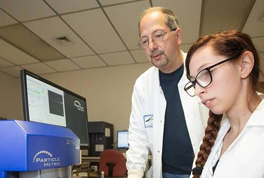 |
| Agilent Seahorse XFe24 Analyzer Installed in the MSB lab in November of 2019, the Seahorse measures the oxygen consumption rate (OCR) and extracellular acidification rate (ECAR) of live cells in a 24-well plate format. Seahorse XF technology measures the flux of oxygen and protons in the transient microchambers created by specially designed tissue culture microplates in real time. The software calculates the rates of oxygen consumption and extracellular acidification from its measurements of oxygen and proton flux. Location: USA COM Medical Sciences Building, Room 2264 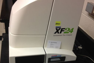 |
| 10x Genomics Chromium iX Controller The newest addition to FCSRLs fleet of instrumentation was installed January 2026. The Chromium iX system employs advanced Next GEM microfluidics to automate partitioning of single cells or nuclei into nanoliter droplets, enhancing reproducibility and reducing user error in single-cell workflows. Its scalable architecture is built on the Chromium X hardware and allows seamless software upgrades for high-throughput capabilities. Optimized for standard throughput, the iX is suitable for everyday experiments and pilot studies, capturing 500 to 20,000 cells per sample (up to 160,000 per run) with up to 80% recovery efficiency. As of 2026, the iX supports standard 10x Genomics assays, including high-sensitivity GEM-X Universal kits. The controller combines a single cell with a single gel bead that is coated with a unique oligonucleotide barcode to form a single cell emulsion droplet, which is referred to as a “GEM” (Gel Bead-in-emulsion). Within each GEM the unique barcoded oligonucleotides are used as primers to generate cDNAs, which can be traced back to cell of origin. The barcoded amplified fragments from hundreds to thousands of cells are pooled to create short read sequencer compatible libraries. Sequencing of these libraries enables analysis of single cells for gene expression, cell surface proteins, immune clonotype, antigen specificity, and chromatin accessibility. There are currently no user fees associated with this system, but advanced scheduling is required. Location: USA COM Medical Sciences Building, Room 2264 |
| Miltenyi Biotech GentleMACS tissue disassociation system The industry standard for preparing single cell suspensions from most any tissue with the highest viability. Location: USA COM Medical Sciences Building, Room 2264 |
| CellRad+ cabinet x-ray irradiator The CellRad+ x-ray system is a compact, tabletop unit featuring an adjustable specimen shelf with a range of 13-44cm (5-17 inches), an electrically operated turntable, and integrated closed-loop cooling. It ensures safety and compliance with US and International regulations for cabinet x-ray systems, requiring no additional shielding. The system includes an integrated dosimeter with Advanced Dose Control and an automatic warm-up function, along with a daily dose QA program to maintain accuracy. User interaction is facilitated by an on-unit, graphical touch-screen interface offering programmable exposure settings and one-touch program recall for streamlined operation. The CellRad+ has a maximum dose rate (based on shelf height) of approximately 8 Gy/minute. Location: Mitchell Cancer Institute (MCI) |
| Kimtron IC-320 X-ray irradiation system The Kimtron IC-320 is a self-contained cabinet X-ray irradiation system designed for precise and repeatable irradiation of biological specimens, including cells and small animals. It serves a broad range of scientific and industrial purposes, primarily focusing on biological research such as irradiating cell cultures, yeast, and small animals for DNA damage, stem cell ablation, and tumor treatment studies. Additionally, the system is frequently employed for the sterilization of various solutions and lab-related model systems, providing a versatile alternative to traditional gamma irradiation. Location: USA COM Medical Sciences Building, Room 1045 | 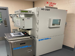 |
| Finesse 325 Microtome Formalin-fixed paraffin embedded (FFPE) tissues can be sectioned using the Finesse 325 microtome (Thermo Scientific) located in the MSB facility. FFPE sections can be examined using various histological and immunohistological approaches. Location: USA COM Medical Sciences Building 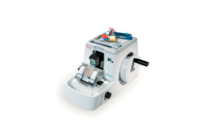 |
Additional instrumentation available for use, but not managed by the Flow Cytometry Shared Resource Laboratories:
- Agilent BioTek Cytation 7 Imaging System (MSB 2264)
The Bioimaging facilities at the USA College of Medicine manage the Agilent BioTek Cytation 7, a versatile imaging system that combines digital upright microscopy, digital inverted microscopy, and multimode microplate detection in one unit. Its inverted widefield module supports fluorescence and brightfield imaging at magnifications ranging from 1.25x to 60x, making it ideal for everything from whole-organism studies to high-resolution intracellular analysis. Complementing this is an upright reflected light module designed for specialized tasks like ELISpot, colony counting, and rapid slide scanning. As a high-performance plate reader it utilizes quadruple monochromator-based optics. It supports multiple detection modes including fluorescence
intensity (top and bottom reading), UV-Vis absorbance (230 nm to 999 nm), luminescence, and time-resolved fluorescence (TRF) and TR-FRET.
For assistance contact:
Joel F. Andrews, Ph.D.
Associate Professor, Research
Department of Biochemistry and Molecular Biology
Manager, BioImaging Core Facility
Frederick P. Whiddon College of Medicine
USA Mitchell Cancer Institute
University of South Alabama
jandrews@southalabama.edu
(251) 410-4927 - Beckman Coulter Multisizer 4e (Earth and Life Sciences Building)
Dr. Nathan Chasen has graciously made his instrument available to other investigators. The Beckman Coulter Multisizer 4e is a high-resolution automated particle analyzer used for counting and characterizing particles and cells. It utilizes the Coulter Principle, also known as electrical sensing zone technology, which measures the change in electrical resistance as particles suspended in a conductive electrolyte pass through a small aperture. The Multisizer 4e provides comprehensive particle and cell analysis using the accurate, "direct" Coulter method, which measures true volume regardless of optical properties. Key specifications include the detection of particles from 200 nm to 1,600 μm, delivering precise data on sample cell number, size, and concentration. The instrument analyzes particles individually, allowing for real-time detection of average cell size changes and identification of individual particles or cell aggregates that deviate from the sample mean.
For assistance contact:
Nathan Chasen, PhD
Assistant Professor of Biology
Department of Biology
ELSB 123
University of South Alabama
nchasen@southalabama.edu
(251) 460-7533