Radiology Sections
Abdominal Imaging
The area of study devoted to abdominal imaging performs diagnostic studies using fluoroscopy, computed tomography (CT scans), ultrasound, and magnetic resonance imaging (MRI).
Computed Tomography
CT scans are performed using advanced medical and multidetector CT scanners with rapid scanning capabilities as well as 3-D reconstruction. CT scans are used for diagnostic treatment of abdominal disorders, angiography, CT-guided biopsies/drainages, catheter placement, tumor detection, and virtual colonography.
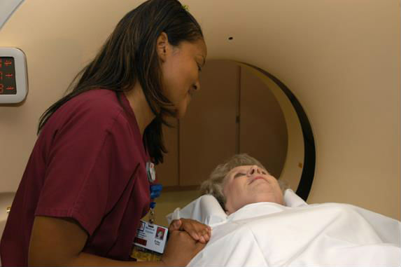
MRI
Magnetic Resonance Imaging is used in the diagnosis of all types of abdominal pathology. In addition, MRI is available for evaluation throughout the body including MRA (angiography) and MRV (venography), both of which provide a less invasive method of screening and detection of vascular abnormalities, such as aneurysms or stenosis.
 Gastrointestinal Imaging
The USA Department of Radiology offers the full spectrum of gastrointestinal imaging from traditional barium studies through to virtual colonoscopy.
Genitourinary Imaging
The genitourinary imaging in the USA Department of Radiology includes complete range of diagnostic procedures with evaluation of the kidneys, bladder, ureters, and gynecological organs using ultrasound, CT, radiography and MRI. CT and ultrasound are used for procedural guidance including for biopsies and drainages.
Breast Imaging
Mammography services recently moved to the new USA Health Strada Patient Care Center, located across from USA Children's & Women's Hospital. In our new location, we continue to give our mammogram patients quick, convenient and thorough treatment. Our Selenia digital mammography system has the capability to detect breast cancer earlier in more women than the film mammography at many hospitals. Breast cancer is the most commonly diagnosed cancer in women, and digital mammography provides an important tool for early detection, which is the best hope for treatment and recovery.
 Cardiothoracic Imaging
In Chest Radiology we use digital radiography, CT scans, and magnetic resonance imaging (MRI) to examine and interpret thoracic pathology. The treatment of chest disease includes interventional diagnostic procedures including needle biopsies as well as lung and abscess drainages.
Musculoskeletal Imaging
The musculoskeletal section of radiology utilizes state-of-the-art imaging including digital radiography, ultrasound, CT and MRI in the diagnosis of trauma, body tumors, arthritis, sports injuries and issues. Additionally, fluoroscopic procedures are performed including arthrograms and joint aspirations/injections. A complete range of procedures are offered in the MRI section at both USA Health University Hospital and USA Children’s & Women’s Hospital, including joint and tumor studies. Both MRA and MRV studies are performed as an alternative to more invasive procedures. MR spectroscopy is used to investigate brain pathology.
Neuroradiology
The neuroradiology section performs all procedures concerning the central nervous system. Radiographs, CT and MRI, including 3-D reconstruction, are all utilized to evaluate the entire central nervous system and brain. Special procedures employed include myelograms, spinal cord angiograms, cerebral angiograms and arteriography of both the internal and external carotid arteries. Interventional neuroradiology procedures include coil and balloon embolizations as well as stroke intervention using MERCI retrieval devices and intra-arterial thrombolysis. A complete range of procedures are offered in the MRI section at both USA Health University Hospital and USA Children’s & Women’s Hospital. Both MRA and MRV studies are performed as an alternative to more invasive procedures. MR spectroscopy is used to investigate brain pathology.
Nuclear Imaging and Therapies / Positron Emission Tomography (PET)
A state-of-the-art positron emission tomography (PET) scanner is used to diagnose and manage all forms of cancer at the USA Mitchell Cancer Institute. We are also leading the way in evaluation of bone metastases with combined scanning with FDG and Na-F PET/CT. The nuclear medicine faculty and staff have extensive expertise in evaluating and managing patients with the full armament of nuclear medicine radiotracers.
Pediatric Imaging
Imaging services at USA Children's & Women's Hospital are tailored to use the concept of "Imaging Gently" to limit exposure in children, deferring to ultrasound and magnetic resonance imaging when possible.
Vascular Imaging & Interventional Radiology
This section provides a wide range of both vascular and non-vascular interventional procedures. Digital subtraction angiography is performed in evaluation of all vascular procedures. Interventional radiology includes routine angiography, venography, balloon angioplasty, placement of intravascular stents, chemoembolization, placement of intra-arterial chemotherapy infusion, catheters, embolization of vascular malformation, tumors, or active bleeding vessels, TIPS procedures, ureteral/biliary dilation and stenting. Abscess drainages and image guided biopsies are also performed in this section utilizing CT and ultrasound.
Interventional Radiology also provides pain relief and management. Currently, the major focus is chronic low back pain management and vertebroplasty. Procedures available for low back pain include epidural, facet joint and sacroiliac joint steroid injections. Radiofrequency ablation of metastatic bone lesions is also used for pain relief.
Ultrasound
Ultrasound imaging at the USA Department of Radiology employs contemporary imaging techniques and scanners, including the new GE Logiq S8 ultrasound scanners at the USA Health University Hospital. The new machines provide state-of-the-art imaging, including elastographic assessment. These new scanners are particularly helpful in assessment of superficial lesions and, in conjunction with the L8-18i-D procedure transducer, biopsy guidance for these lesions.